Follow the reproductive cells, then locate each event
Human reproduction is easier to revise as a sequence of locations and cell events than as two disconnected anatomy lists. The broad route is gamete formation, transfer of sperm into the female reproductive tract, fertilisation, early development, implantation and placental connection. A structure matters because it enables one part of that route.
Keep gamete formation distinct from fertilisation. Spermatogenesis produces sperm in the testes. Oogenesis begins in the ovaries, but the cell released at ovulation is a secondary oocyte, arrested in metaphase II. Sperm entry triggers completion of that division. Fertilisation normally occurs at the ampullary-isthmic junction of the oviduct; fusion of the haploid nuclei establishes the diploid zygote.
From zygote to implantation: sequence before terminology
After fertilisation, the zygote undergoes cleavage while moving toward the uterus. The early dividing cells form a morula, and a later blastocyst has an outer trophoblast layer and an inner cell mass. The terms are related by sequence, not interchangeable labels for the same stage.
Implantation occurs when the blastocyst becomes embedded in the uterine endometrium. Do not place implantation in the oviduct merely because fertilisation happens there. A reliable location check is: fertilisation in the oviduct, then early divisions during transit, then implantation in the uterus.
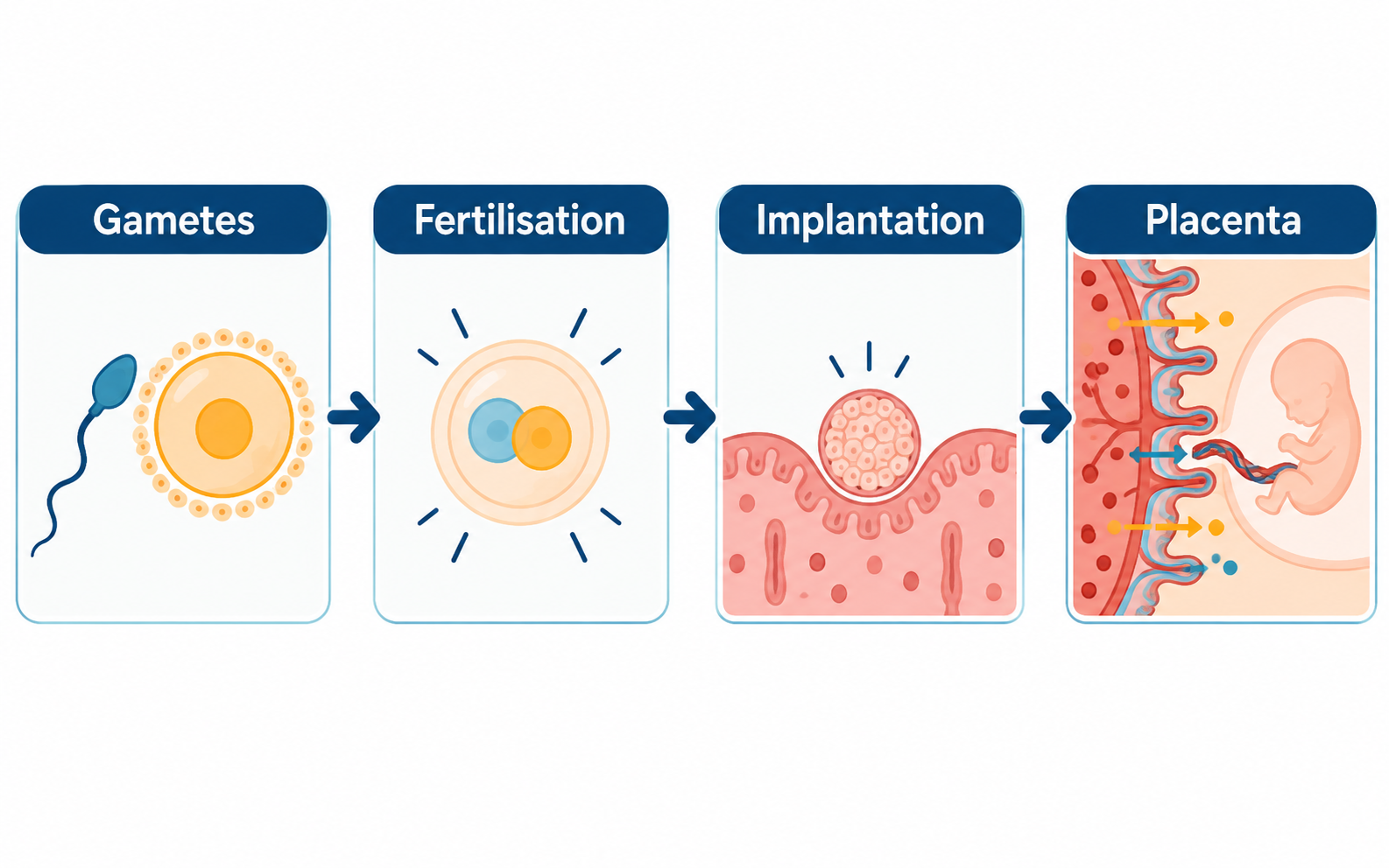
The place-before-stage check
Attach every term to a location before you memorise a sequence. A cell event, a developmental stage and an anatomical structure are not interchangeable answers.
- Gametes are produced in gonads; a zygote forms only when sperm and ovum fuse.
- Fertilisation is normally described in the ampullary-isthmic region of the oviduct.
- The blastocyst implants in the uterine endometrium after early cleavage stages.
- The placenta is an exchange interface; the umbilical cord connects the developing foetus to it.
A route is more useful than a crowded anatomical drawing
The most common sequence error is to group fertilisation, cleavage and implantation under the uterus because the later stages occur there. Keeping the oviduct-to-uterus transition visible makes the stages easier to place.
Use the route to test every unfamiliar word: does it name a cell, a developmental stage, a place or an exchange structure? Those categories cannot occupy the same position in the sequence.
Human reproduction: event, location and direct outcome
Follow the cell event through its location. This prevents fertilisation, implantation and placental exchange from being placed in the same structure.
| Biological event | Main location | Direct outcome | Do not swap with |
|---|---|---|---|
| Gamete formation | Testes or ovaries | Sperm or ova are produced | Fertilisation |
| Fertilisation | Ampullary-isthmic region of oviduct | Zygote forms after gamete fusion | Implantation in uterus |
| Implantation | Uterine endometrium | Blastocyst becomes embedded | Early cleavage during transit |
| Placental exchange | Placenta | Maternal and foetal exchange interface | Umbilical cord, which connects foetus and placenta |
Placenta links two circulations without mixing them directly
The placenta forms an exchange interface between the developing foetus and the pregnant person's body. It supports transfer of materials such as nutrients and gases and is associated with hormone production during pregnancy. The revision point is the interface: maternal and foetal blood do not simply become one circulating pool.
The umbilical cord connects the developing foetus with the placenta. In a route question, distinguish the cord as the connection from the placenta as the exchange surface. The amnion and amniotic fluid provide a protective environment around the developing foetus; they are not substitutes for the placenta's transport role.
Count the products of meiosis, not just the divisions
One primary spermatocyte completes meiosis I to give two secondary spermatocytes; meiosis II produces four haploid spermatids. Their transformation into sperm is spermiogenesis. Release from Sertoli cells into the tubule lumen is spermiation. These last two events describe different actions and neither is another chromosome-reduction division.
Oogenesis distributes the cytoplasm unequally. A primary oocyte begins meiosis before birth and arrests in prophase I. Completion of meiosis I produces a large secondary oocyte and a much smaller polar body. Keeping most of the cytoplasm in one cell supports early development. The parallel with four equally sized spermatids therefore breaks down even though both pathways involve meiosis.
As an original counting exercise, start with three primary spermatocytes. Assuming every meiotic product survives, they yield twelve spermatids, each haploid. Three primary oocytes do not yield twelve functional ova. Explain the unequal cytoplasmic division before trying to count the polar bodies.
Read the ovarian cycle alongside the uterine cycle
FSH and LH come from the anterior pituitary, whereas developing ovarian follicles produce oestrogens. During the follicular phase, follicle development is accompanied by rebuilding of the endometrium. Sustained high oestrogen near mid-cycle contributes to positive feedback and the LH surge that triggers ovulation. An ovulation arrow belongs at the follicle, not at the uterine lining.
After ovulation the ruptured follicle forms the corpus luteum, which secretes progesterone and supports a secretory endometrium. In a cycle without pregnancy, corpus-luteum regression lowers ovarian hormone levels and the lining is shed. During early pregnancy, hCG supports the corpus luteum. A textbook 28-day diagram is a model for understanding the sequence, not a universal timetable or a way to predict an individual's fertility.
Cleavage increases cell number before it increases embryo size
During early cleavage, repeated mitotic divisions partition the zygote into smaller blastomeres. The total structure does not double in size each time a cell divides. A 16-cell morula is not sixteen full-sized zygotes joined together. Later, fluid accumulation helps establish the blastocyst cavity and separates the outer trophoblast from the inner cell mass.
Test the route with three statements: a secondary oocyte is ovulated; a blastocyst implants; the placenta provides an exchange interface. Replacing any of those stages with 'egg' hides a different biological event. At birth, uterine contractions involve positive feedback with oxytocin. After birth, prolactin supports milk production while oxytocin supports milk ejection: production and movement are separate functions.
A location-and-stage recall map
Draw three place labels in a row: gonad, oviduct and uterus. Under them, place gamete formation, fertilisation and implantation respectively. Add one final side box for placenta and join it to the developing foetus with a cord. This four-place map is enough to check most sequence errors without copying a detailed anatomical diagram.
Finish with three verbal checks: Which cells fuse at fertilisation? Which stage implants? What is the difference between a placenta and an umbilical cord? The answer should name both the event and its location.
Common confusions to check
- A gamete is not a zygote; a zygote forms only after sperm and ovum fuse.
- Fertilisation is normally described in the oviduct, while implantation occurs in the uterine endometrium.
- Placenta and umbilical cord have linked but different roles: exchange surface versus connection.
Editorial note and disclaimer
Written by: DongFeng. Published by: MedQGo. Last updated: September 18, 2026.
Disclaimer: This guide is a revision aid for NEET-UG aspirants and does not constitute medical advice. For clinical or health-related queries, consult a qualified medical professional.
References
Related revision guides
How to use this guide
Read the relevant NCERT chapter first. Then redraw the relationships or process described here from memory, compare your version with the textbook, and correct only the gaps. This is an independent revision aid, not official NCERT, NTA, or NEET material.