Use disorder examples to revise inheritance, not diagnosis
NCERT introduces named genetic disorders to show how altered genes or chromosome number can be inherited or detected through family and chromosome patterns. For this revision guide, keep the scope educational: the aim is to identify the inheritance category and the evidence used for classification. It is not a guide for diagnosis, treatment or personal risk assessment.
The first split is between Mendelian disorders and chromosomal disorders. Mendelian disorders are mainly determined by alteration in a single gene and can be traced through pedigree analysis. Chromosomal disorders involve absence, excess or abnormal arrangement of one or more chromosomes, often visible through karyotype-level reasoning. Mixing those two categories is the biggest avoidable error.
Autosomal recessive examples need both copies in the model
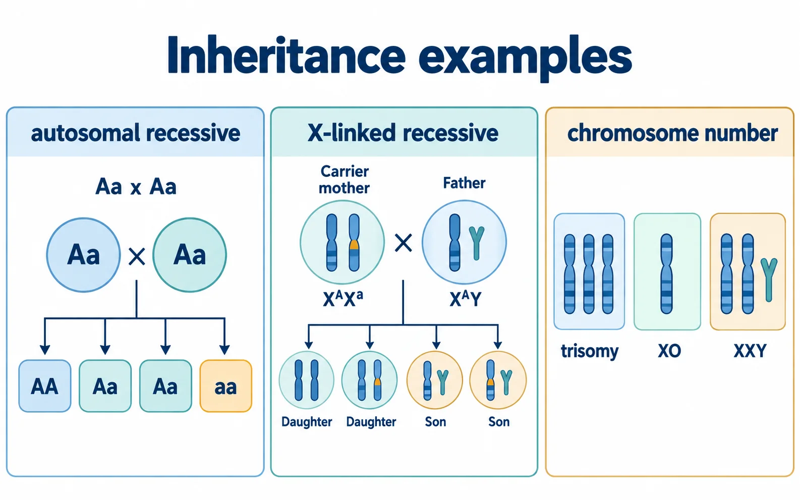
In an autosomal recessive model, the affected phenotype is usually shown when both copies of the gene carry the relevant altered allele. Two unaffected carrier parents can produce unaffected non-carrier, carrier and affected offspring in a Punnett-square model. The familiar 1:2:1 genotype pattern belongs to the stated carrier-cross model, not to every family in the world.
NCERT uses sickle-cell anaemia, phenylketonuria and thalassaemia as examples within the broader discussion of altered genes. For revision, focus on the inheritance logic and the specific molecular point only where NCERT states it. Sickle-cell anaemia is described as an autosome-linked recessive trait involving the haemoglobin beta chain; NCERT also contrasts it with thalassaemia by noting qualitative versus quantitative globin problems. Phenylketonuria is treated as an autosomal recessive inborn error involving phenylalanine metabolism.
A safe answer does not turn those facts into personal health advice. It says what kind of inheritance example is being used and what evidence supports the category.
A health example is still a genetics exercise here
Named conditions can make a revision page feel clinical, but the chapter is using them to teach inheritance. Keep the answer at that level unless a qualified medical context is explicitly involved. In a study note, the honest work is to identify the route of inheritance, the evidence and the boundary of what the textbook statement supports.
Genetic disorder examples: choose the evidence scale first
The names are useful only after the inheritance scale is clear. Start from allele route or chromosome count, then add the example.
| Category | NCERT example | Evidence scale | Common trap |
|---|---|---|---|
| Autosomal recessive | Sickle-cell anaemia, phenylketonuria, thalassaemia | Two altered gene copies or carrier-cross logic | Treating a carrier as affected |
| X-linked recessive | Colour blindness, haemophilia | Sex-chromosome route through X contribution | Assuming father-to-son X-linked transmission |
| Autosomal dominant | Example category in pedigree analysis | Affected pattern across generations | Calling every pedigree recessive |
| Chromosomal disorder | Down, Turner and Klinefelter syndromes | Aneuploidy or sex-chromosome count | Forcing a karyotype clue into a single-gene cross |
X-linked recessive examples change the transmission route
X-linked recessive inheritance cannot be read like an autosomal recessive cross because males and females do not contribute sex chromosomes in the same way. A father passes his X chromosome to daughters and his Y chromosome to sons. A carrier mother can pass either X chromosome to each child. That route explains why some X-linked recessive traits are more commonly expressed in males.
Colour blindness and haemophilia are used as sex-linked recessive examples in the NCERT inheritance chapter. The key revision point is not the symptom detail. The key point is chromosome route: the altered allele is linked with the X chromosome, and expression depends on which sex chromosome combination the offspring receives.
Before choosing an option, write the route. Mother: X with normal allele or X with altered allele. Father: X or Y. Then ask which offspring class can express the recessive allele without a second X carrying a normal copy. This route check prevents the common father-to-son X-linked error.
Chromosomal disorders are number or arrangement problems
Chromosomal disorders sit at a different biological scale. Instead of one gene allele, the issue is an extra, missing or rearranged chromosome segment or chromosome. NCERT highlights aneuploidy as gain or loss of chromosome number following failure of proper segregation, and notes polyploidy as an increase in the whole set of chromosomes, often seen in plants.
Down syndrome is presented as trisomy of chromosome 21. Turner syndrome is linked with loss of one X chromosome, and Klinefelter syndrome with an XXY karyotype. The revision task is to recognise that these are chromosome-number examples, not simple single-gene recessive crosses.
That distinction matters in MCQs. A pedigree-style allele cross is useful for a Mendelian example; a karyotype-style count is useful for a chromosomal disorder. If the evidence names 47 chromosomes, trisomy, XO or XXY, start from chromosome number before trying to assign a single-gene genotype.
Worked example: decide the evidence type first
Consider three statements. Statement A says an unaffected couple are both carriers and one child expresses an autosomal recessive trait. Statement B says a carrier mother can transmit an X-linked recessive allele to a son. Statement C says an individual has an extra chromosome 21. These statements do not belong to the same reasoning tool.
A is a Mendelian autosomal recessive model, so a Punnett square with two carrier genotypes is appropriate. B is an X-linked recessive route, so the useful drawing is a sex-chromosome contribution map. C is a chromosomal-number case, so the useful evidence is a karyotype-style count. The mistake would be to force all three into one generic 'genetic disease' table.
This is the habit to practise: name the scale first. Gene allele, sex-linked allele or chromosome number. Once the scale is correct, the example becomes much less confusing.
From a carrier cross to a checkable probability
Now work through statement A using an invented single-gene model. Let A be the dominant allele and a the recessive allele; assume AA and Aa are unaffected and aa expresses the trait. For Aa x Aa, each parent produces A and a gametes with equal probability. Combining the gametes gives AA, Aa, aA and aa, each with probability 1/4. Aa and aA describe the same heterozygous genotype, so the genotype ratio is 1 AA : 2 Aa : 1 aa.
The result is a 1/4 probability of expressing the trait, a 1/2 probability of being an unaffected carrier and a 1/4 probability of being an unaffected non-carrier at each fertilisation under these assumptions. Four boxes are four equally likely combinations, not a prediction that four children must include one affected child. Each new fertilisation is another event; the model does not balance earlier outcomes.
Check a plausible wrong answer: 'Half of the unaffected offspring are carriers.' The denominator has changed. Among the three unaffected combinations, AA, Aa and aA, two are carriers, so the conditional probability is 2/3, not 1/2. Use 1/2 when considering all offspring; use 2/3 when the model additionally tells you the offspring is unaffected. These are classroom-model probabilities, not an assessment of any person's health.
Comparison table before memorising names
The table below keeps each named example tied to the kind of evidence that supports it. Do not use it as a medical list. Use it as a genetics filter: what changed, where the evidence is read and what trap the option may set.
Common confusion checks
- Mendelian disorder does not mean every case is autosomal; haemophilia and colour blindness are sex-linked recessive examples in NCERT.
- Carrier status and affected status are different categories in recessive inheritance.
- Sickle-cell anaemia and thalassaemia are both linked with haemoglobin, but NCERT distinguishes qualitative and quantitative globin problems.
- Down syndrome, Turner syndrome and Klinefelter syndrome are chromosome-number examples, not ordinary single-gene Punnett-square examples.
- A revision note can explain inheritance categories, but personal medical questions require a qualified professional.
A 15-minute recall routine
Draw three boxes: autosomal recessive, X-linked recessive and chromosomal number. Put two NCERT examples in the first box, two in the second and three chromosome-number examples in the third. Then write the evidence beside each box: carrier cross, sex-chromosome route or karyotype count.
End with one spoken explanation: 'I would not test these examples with the same diagram because the biological scale is different.' That sentence is the heart of the guide.
Common confusions to check
- Carrier and affected status are not the same in recessive inheritance.
- An X-linked trait is not transmitted father to son through the X chromosome.
- Down, Turner and Klinefelter examples are not ordinary single-gene crosses.
Editorial note and disclaimer
Written by: DongFeng. Published by: MedQGo. Last updated: September 29, 2026.
Disclaimer: This guide is a revision aid for NEET-UG aspirants and does not constitute medical advice. For clinical or health-related queries, consult a qualified medical professional.
References
Related revision guides
How to use this guide
Read the relevant NCERT chapter first. Then redraw the relationships or process described here from memory, compare your version with the textbook, and correct only the gaps. This is an independent revision aid, not official NCERT, NTA, or NEET material.