A valve responds to pressure, not to a memorised phase name
The circulation route tells you where blood goes; the cardiac cycle explains when it can move between compartments. Start with three places on one side of the heart: atrium, ventricle and outgoing artery. A valve between two of them responds to the pressure difference across it. Knowing only that a chamber is contracting is not enough to decide whether its outlet has opened.
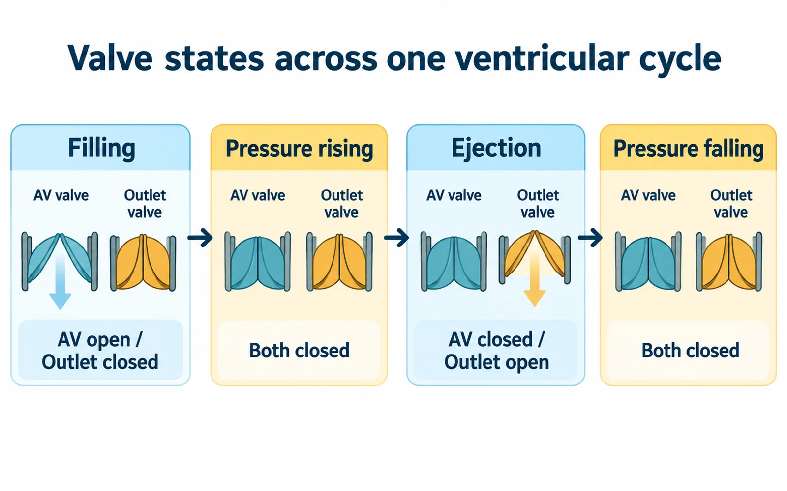
Use the left side as a reference: the mitral valve lies between left atrium and ventricle, and the aortic valve guards the outlet. The corresponding right-sided valves are tricuspid and pulmonary. Both sides follow the same broad order of events, although their pressures differ. In the illustration, AV means atrioventricular and outlet means semilunar; the symbols are functional gates, not anatomical drawings.
Four states make the closed-valve intervals visible
During ventricular filling, atrial pressure exceeds ventricular pressure sufficiently to open the AV valve. The semilunar valve remains closed. Much filling occurs before the atrium contracts; atrial systole adds to it. Ventricular contraction then raises pressure, closing the AV valve. There is a short interval before ventricular pressure exceeds arterial pressure, so the outlet is still closed too.
That early interval is isovolumetric contraction: pressure rises while the enclosed ventricular blood volume stays approximately constant. Once ventricular pressure exceeds the outgoing artery's pressure, the semilunar valve opens and ejection occurs. The AV valve stays closed, preventing a route back towards the atrium.
As the ventricle relaxes, pressure falls below arterial pressure and the semilunar valve closes. Before it falls sufficiently below atrial pressure to reopen the AV valve, both valves are closed again. This is isovolumetric relaxation. Filling resumes after the AV valve opens. Read the four states as a sequence of pressure crossings, not four equal time blocks.
Closed does not mean motionless
With both valves closed, the ventricular wall can still change tension and pressure. Approximately constant blood volume is the feature named by isovolumetric, not an absence of muscle activity. State what is staying constant before interpreting the phase.
Read valve state alongside the volume change
This idealised ventricular sequence assumes competent valves. Both-closed states occur twice, so pressure direction is needed to distinguish them.
| Ventricular interval | AV valve | Semilunar valve | Ventricular volume |
|---|---|---|---|
| Filling | Open | Closed | Increases |
| Isovolumetric contraction | Closed | Closed | Approximately constant while pressure rises |
| Ejection | Closed | Open | Decreases |
| Isovolumetric relaxation | Closed | Closed | Approximately constant while pressure falls |
Solve three pressure snapshots before naming their phases
Consider an invented idealised left-heart model. Use arbitrary pressure units solely to compare compartments; these are not physiological reference values or clinical measurements. Assume competent valves that respond promptly to the pressure differences. In snapshot A, atrial pressure is 8, ventricular pressure 5 and arterial pressure 70. The atrium can drive flow into the ventricle, while the artery-to-ventricle gradient holds the outlet closed.
In snapshot B, keep atrial pressure at 8 and arterial pressure at 70, but let ventricular pressure rise to 25. The ventricle now exceeds the atrium but not the artery. The AV valve is closed and the outlet has not opened: both are closed. This is compatible with the rising-pressure interval before ejection. Without the stated rising trend, the three numbers alone would not establish whether the ventricle was contracting or relaxing.
In snapshot C, ventricular pressure reaches 85 while atrial and arterial values remain 8 and 70. The outlet can now open, and the AV valve remains closed. The wrong answer 'the outlet opens as soon as ventricular pressure exceeds atrial pressure' compares the wrong pair. Each valve has its own two-sided comparison.
To reverse the reasoning, imagine pressure falling after ejection through a value between arterial and atrial pressures. Both valves can again be closed, this time during relaxation. Identical valve states therefore do not uniquely identify the direction of pressure change. Add sequence information before deciding between the two isovolumetric intervals.
Volume change and output are separate accounts
End-diastolic volume is the ventricular volume after filling; end-systolic volume is the amount remaining after ejection. Their difference is stroke volume. In an invented arithmetic example, take 125 mL at the end of filling and 55 mL after ejection. Stroke volume is 125 - 55 = 70 mL per beat. The ventricle has not emptied completely.
If the model heart rate is 75 beats per minute, cardiac output is 70 x 75 = 5,250 mL per minute, or 5.25 litres per minute, for one ventricle. Check the units: mL per beat multiplied by beats per minute leaves mL per minute. A 75-beat rate also gives an average cycle duration of 60/75 = 0.8 seconds. This does not specify how that time is divided among the phases.
Do not add left and right outputs to claim 10.5 litres of fresh systemic flow per minute. The pulmonary and systemic circuits are connected in series, and their average outputs match in a steady-state model. The same circulating blood passes through both pumps. Likewise, the 55 mL residual volume is not another amount to subtract from cardiac output after stroke volume has already been calculated.
A higher rate alone does not guarantee a larger output in an arbitrary comparison. If a second hypothetical state has 100 beats per minute and 45 mL per beat, its output is 4.5 litres per minute. This is arithmetic using supplied values, not a claim about a particular person or a prescription for exercise. The two factors have to be considered together.
Electrical activity, valve sounds and movement need different labels
The sinoatrial node normally initiates the electrical sequence, which spreads through the atria and then the ventricular conduction pathway. Electrical activation precedes the associated mechanical response. An ECG records electrical activity; it is not a plot of ventricular pressure or the amount of blood being ejected.
At the NCERT level, P represents atrial depolarisation, QRS ventricular depolarisation and T ventricular repolarisation. Do not rename QRS 'the outlet-opening wave'. Pressure must still rise sufficiently for ejection after activation. The first and second heart sounds are associated respectively with AV-valve closure and semilunar-valve closure, rather than the sound of those valves opening.
This distinction also helps interpret the word systole. Specify atrial or ventricular systole rather than assuming all four chambers contract together. The guide is about normal sequence reasoning; recognising an abnormal tracing or evaluating symptoms requires clinical assessment and is outside its scope.
Rebuild the cycle from two questions at each boundary
Draw atrium, ventricle and artery in a row. At each valve ask which side currently has higher pressure. Then write the four states from memory: filling, closed-valve pressure rise, ejection, closed-valve pressure fall. Beneath each, mark whether ventricular volume increases, stays constant or decreases. Add atrial contraction to late filling, rather than making it the only source of filling.
Finally solve snapshot B twice: once with pressure rising and once with pressure falling. Both answers have closed valves, but different phase names. Check the sequence against NCERT Class 11 Biology, Chapter 15, section 15.3.2, and distinguish the electrical account in section 15.3.3. The output calculation should finish with units and one ventricle as its reference.
Common confusions to check
- Both valves are closed during two different ventricular intervals.
- An ECG represents electrical activity, not a pressure trace.
- Stroke volume excludes blood remaining after ejection.
- Left and right outputs are not added as independent systemic supplies.
Editorial note and disclaimer
Written by: DongFeng. Published by: MedQGo. Last updated: October 8, 2026.
Disclaimer: This guide is a revision aid for NEET-UG aspirants and does not constitute medical advice. For clinical or health-related queries, consult a qualified medical professional.
References
Related revision guides
How to use this guide
Read the relevant NCERT chapter first. Then redraw the relationships or process described here from memory, compare your version with the textbook, and correct only the gaps. This is an independent revision aid, not official NCERT, NTA, or NEET material.