Use the body cavity as a map, not a list
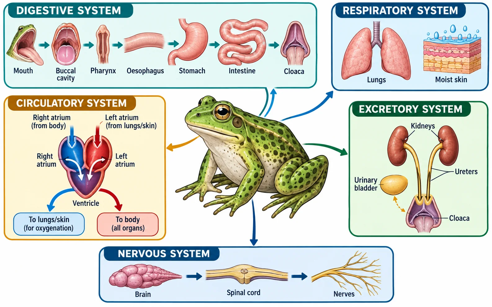
A frog's systems are easier to revise when each is followed from an entry point to an exit or destination. Food travels through a continuous canal; blood moves through a closed circulation; air exchange uses both lungs and skin; urine reaches the cloaca through the urinary route; and neural signals travel from the brain through the spinal cord and nerves. These routes occupy the same animal but answer different questions.
This guide follows the NCERT account of an adult frog as a structural-organisation example. The 2026 NEET syllabus lists brief anatomy and functions of the digestive, circulatory, respiratory, nervous and reproductive systems of an insect (frog), preserving the syllabus wording. The scope here is the frog model described in NCERT, not a dissection protocol or wildlife-care advice.
Food follows one canal before wastes leave through the cloaca
The digestive route begins at the mouth and continues through the buccal cavity, pharynx, oesophagus, stomach, intestine and rectum to the cloaca. Digestive glands contribute secretions: the liver produces bile, stored in the gall bladder, while the pancreas produces pancreatic juice. The alimentary canal is the route taken by food; a gland can aid digestion without being a segment through which food passes.
The NCERT text relates the comparatively short intestine to the frog's carnivorous diet. Keep that explanation attached to the stated model rather than turning it into a universal rule for every carnivore. A useful tracing task is to underline the canal organs in one colour and circle the accessory glands in another. If the liver appears in the food route, the map has confused secretion with passage.
In a simple invented route check, a labelled arrow runs mouth -> oesophagus -> stomach -> intestine -> cloaca but omits the buccal cavity and pharynx. The final destination is plausible, yet the route is incomplete. Restore the missing intermediate structures before describing where digestion or absorption occurs. This checks sequence, not a clinical or experimental procedure.
A shared chamber does not merge the routes
The cloaca receives material from more than one system, but each route has a distinct origin and function. Trace backward from the outlet before naming the system: intestine points to digestion, while ureters point to the urinary tract.
The circulatory diagram is intentionally simplified. Use the chamber count and major routes for revision; do not infer clinical significance or detailed flow measurements from it.
Frog systems: route, structure and common mix-up
Trace each system independently even when two routes meet at the cloaca.
| System | Key structure or route | Functional checkpoint | Do not confuse |
|---|---|---|---|
| Digestive | Mouth to buccal cavity, pharynx, oesophagus, stomach, intestine, rectum, cloaca | Liver and pancreas add secretions | Accessory glands are not food-canal segments |
| Circulatory | Two atria and one ventricle; pulmocutaneous and systemic routes | One ventricle receives blood from both atria | Double circulation does not mean four chambers |
| Respiratory | Moist skin, lungs and buccopharyngeal lining | Gas exchange occurs at respiratory surfaces | Gas exchange is not ventilation or blood transport |
| Urinary | Kidneys to ureters to cloaca; bladder stores urine | Kidneys form urine | Cloaca is an outlet, not the urine-forming organ |
| Nervous | Brain to spinal cord to peripheral nerves | Carries and coordinates signals | A signal route is not a fluid route |
One ventricle serves a three-chambered heart
The adult frog heart has two atria and one ventricle. The right atrium receives deoxygenated blood returning from the body; the left atrium receives oxygenated blood from the lungs and skin. Both atria empty into the single ventricle. Unlike the four-chambered human heart, the ventricle is not divided into separate right and left chambers, so do not draw two ventricular cavities.
Frogs have a double circulation in the broad sense that blood passes through the heart on its way through the body and the respiratory surfaces. Pulmocutaneous circulation links the heart with lungs and skin, while systemic circulation supplies the body. Because the ventricle is single, the routes are not anatomically separated there as in a four-chambered mammalian heart. The diagram is a route map, not a detailed account of every flow pattern or mixing event.
A common incorrect answer calls the frog heart 'two-chambered' because it has one ventricle. Count all chambers: two atria plus one ventricle equals three. Another shortcut assumes that every double circulation requires four chambers. Here, name the two circulation routes and separately state the chamber arrangement.
Gas exchange uses skin as well as lungs
The respiratory account should include both cutaneous respiration through the moist skin and pulmonary respiration through the lungs. The skin's role is not a decorative add-on to the lung route. It is a respiratory surface, which is why moisture matters to gas exchange. Frogs can also exchange gases through the buccopharyngeal lining in the NCERT account; the relative contribution depends on the context described.
Keep three terms apart: ventilation moves air, exchange moves gases across a respiratory surface, and circulation transports gases in blood. A lung diagram alone cannot represent all frog respiration. Conversely, saying 'frogs breathe through skin' does not make lungs irrelevant. The question may be asking for a surface, a movement of air or a transport route; identify which level is requested before answering.
Kidneys, urinary passages and reproductive ducts meet at a shared outlet
The kidneys filter blood and form urine. Urine passes through the ureters toward the cloaca; the urinary bladder can store it before elimination. The cloaca is a common chamber that receives products from more than one system, but it does not make the digestive, urinary and reproductive pathways identical. Follow each route to the shared region, then name its origin.
Frog reproduction is sexual, with separate sexes in the standard NCERT description. Eggs and sperm are released into water, where fertilisation is external. The reproductive passage and urinary passage have sex-specific anatomical relations, so a single simplified diagram should not be used to infer every duct in both sexes. For a short recall answer, state the process (external fertilisation), the setting (water) and the gamete route at the level required by the text.
A likely mix-up is 'the cloaca produces urine'. It is an outlet chamber, not the kidney. Write the route as kidney -> ureter -> cloaca, with the bladder as a storage connection, and separately trace digestive contents from the intestine. The shared endpoint does not erase the distinct upstream organs.
The nervous route coordinates rather than transporting material
The brain and spinal cord form the central nervous system; nerves connect it with the rest of the body. A stimulus is detected, information is integrated, and an appropriate response is coordinated through neural pathways. This is a signal route, not a fluid pathway like blood or urine. The distinction matters when the same body map contains several kinds of arrows.
For active recall, draw five labelled lanes: food, blood, respiratory exchange, urine, and neural signalling. Add the start, key intermediate structures and destination for each. Then add one sentence explaining why the cloaca is shared but the canals remain distinct. Compare the heart route with [blood and circulation](/neet-ug/biology/blood-and-circulation), review tissue roles in [structural organisation in animals](/neet-ug/biology/structural-organisation-in-animals), and use [animal kingdom](/neet-ug/biology/animal-kingdom) for the broader chordate context.
A quick route audit
- Food route: mouth -> buccal cavity -> pharynx -> oesophagus -> stomach -> intestine -> rectum -> cloaca.
- Heart: two atria and one ventricle; distinguish pulmocutaneous from systemic circulation.
- Respiration: include skin and lungs; distinguish gas exchange from blood transport.
- Urinary route: kidneys -> ureters -> cloaca; the bladder stores urine.
- Reproduction: separate sexes and external fertilisation in water in the NCERT model.
A 12-minute redraw
Spend four minutes drawing the five lanes from memory, four minutes checking the route endpoints against NCERT, and four minutes correcting only the arrows or organ roles you missed. Finish by explaining aloud why the liver is not part of the food canal, why the frog heart is not two-chambered, and why skin belongs in the respiratory answer. NCERT anchor: Class 11 Biology, Chapter 7, Structural Organisation in Animals, section 7.2, Reprint 2026-27.
Common confusions to check
- The adult frog heart has three chambers, not two or four.
- The cloaca is a shared chamber; kidneys form urine and the intestine carries digested material.
- Frog respiration is not lung-only: moist skin and buccopharyngeal exchange also matter in the NCERT account.
Worked route check: which system reaches the cloaca?
An invented diagram shows one arrow from kidney to ureter to cloaca and another from intestine through rectum to cloaca. Both arrows end at the same chamber, but they do not name the same process: the first is urinary and the second is digestive. The kidneys form urine; the cloaca receives it. The intestinal route carries digestive contents to the outlet.
The tempting wrong answer is 'the cloaca is part of the kidney because urine enters it'. Correct the claim by naming the origin, intermediate route and shared endpoint separately. Then check the heart: two atria plus one ventricle gives three chambers, while the lungs and moist skin provide respiratory surfaces. NCERT anchor: Class 11 Biology, Chapter 7, section 7.2, Structural Organisation in Animals.
Editorial note and disclaimer
Written by: DongFeng. Published by: MedQGo. Last updated: October 10, 2026.
Disclaimer: This guide is a revision aid for NEET-UG aspirants and does not constitute medical advice. For clinical or health-related queries, consult a qualified medical professional.
References
Related revision guides
How to use this guide
Read the relevant NCERT chapter first. Then redraw the relationships or process described here from memory, compare your version with the textbook, and correct only the gaps. This is an independent revision aid, not official NCERT, NTA, or NEET material.