A tissue is not a small organ
A group of similar cells, together with intercellular material where relevant, forms a tissue. An organ contains more than one tissue arranged for a particular job. An organ system coordinates multiple organs. This hierarchy is simple to state but easy to blur when a question names a familiar structure such as skin, stomach or a blood vessel.
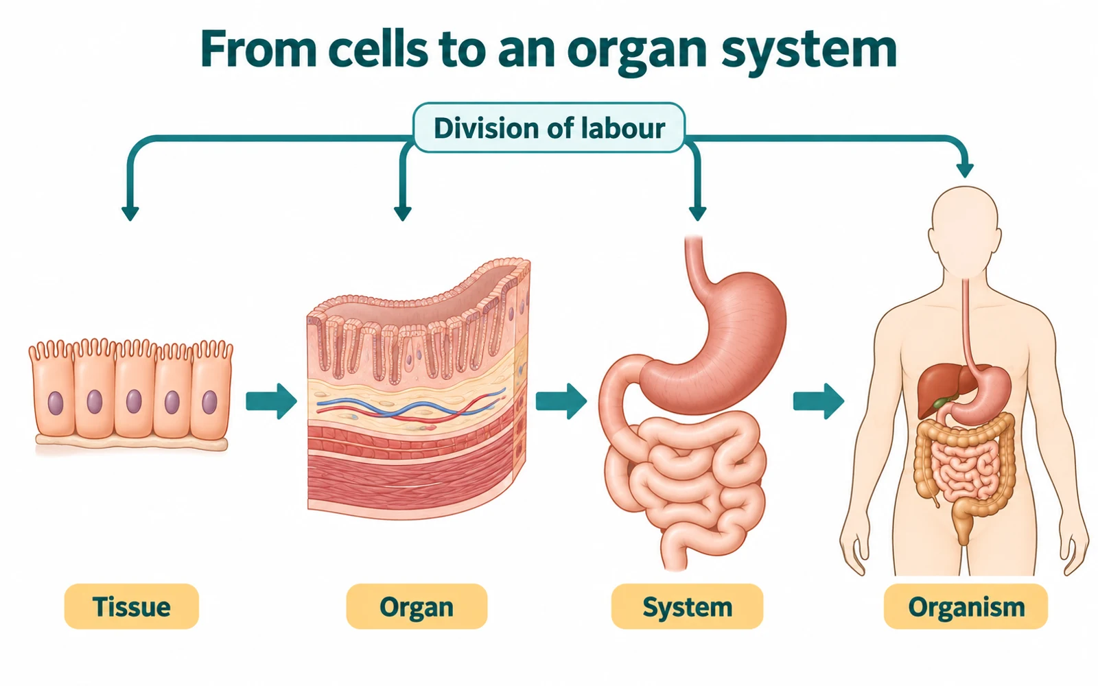
Instead of memorising a list from largest to smallest, trace what changes at each step. Cells specialise; similar cells contribute a shared role; different tissues build a working organ; organs cooperate across a route. The purpose is division of labour, not merely an increase in physical size.
Read the hierarchy as a change in composition
An epithelial sheet is a tissue because its similar cells form a covering or lining. A piece of intestinal wall is an organ because epithelial, connective, muscular and nervous components are arranged together. The digestive system is an organ system because multiple organs coordinate movement, secretion, digestion and absorption. An organism contains many interacting systems.
The illustration uses a digestive example to show that an organ does not arise by enlarging one cell type. It is assembled from multiple tissues. The image is a conceptual hierarchy rather than a detailed anatomical map. A learner should be able to replace the digestive example with a different system while preserving the relationships.
Use arrows for tendon and ligament
Write muscle -> tendon -> bone, then bone -> ligament -> bone. This tests attachment directly, rather than relying on a vague memory that both are strong connective tissues.
Four tissues, four different clues
Use the structural clue and the job together; a location by itself is rarely enough.
| Tissue category | Structural clue | Main role | Distinguish it from |
|---|---|---|---|
| Epithelial | Closely packed cells forming a sheet | Covering, lining or secretion | Connective tissue with abundant matrix |
| Connective | Cells within variable extracellular matrix | Binding, support or transport | Epithelial covering |
| Muscle | Elongated contractile cells | Contraction and movement | Neuron signal transmission |
| Nervous | Neurons with supporting glia | Communication and coordination | Muscle fibre contraction |
Epithelial tissue is defined by position and arrangement
Epithelial tissues cover external surfaces and line internal spaces. Their cells are closely packed with little intercellular material, creating a continuous layer. The shape and number of layers are linked to location and function: simple squamous epithelium provides a thin lining, cuboidal and columnar forms occur in different lining or secretory roles, and stratified epithelium provides protection where wear is greater.
Glandular epithelium specialises in secretion. It should not be called connective tissue simply because its product may enter a fluid or support another organ. The defining question is whether the cells form a covering, lining or secretory epithelial arrangement, not whether the named organ is inside the body.
A plausible wrong shortcut is 'all thin epithelium is for absorption'. Thin sheets can support exchange or lining functions depending on location. Match tissue form and placement before assigning a single verb.
Connective tissue depends on its matrix
Connective tissues connect, support, bind or transport, and their cells are set in varying amounts of intercellular matrix. Areolar tissue fills spaces, adipose tissue stores fat, tendon joins muscle to bone, and ligament joins bone to bone. The last pair is often reversed because both are strong connective tissues; their attachments are the decisive difference.
Cartilage provides flexible support and differs from bone, whose matrix is hard and mineralised. Blood is a fluid connective tissue: formed elements are suspended in plasma, and its transport role does not remove it from the connective category. Calling blood 'only a liquid' overlooks both its cells and its tissue-level role.
For a checkable contrast, write two arrows: muscle -> tendon -> bone and bone -> ligament -> bone. If either arrow starts or ends with the wrong structure, correct the attachment before adding details about elasticity.
Muscle tissue shortens, but its types do not share the same control
Muscle tissue is specialised for contraction. Skeletal muscle is striated and usually voluntary; smooth muscle lacks striations and is involuntary; cardiac muscle is striated and involuntary. The combination matters. 'Striated' alone cannot identify skeletal muscle because cardiac muscle is also striated.
The cells also differ in organisation. Skeletal fibres are long and multinucleate; cardiac cells branch and connect through specialised junctions; smooth muscle cells are spindle-shaped. In an introductory tissue guide, these shape cues support classification, but they do not replace the functional distinction between control and location.
Link this section to locomotion and movement for the sliding-filament mechanism in skeletal muscle. Tissue classification tells you what type of muscle is present; it does not by itself explain every movement of a joint.
Nervous tissue coordinates rapid communication
Nervous tissue contains neurons, specialised for receiving and transmitting signals, and supporting neuroglial cells. A neuron has a cell body, dendrites that commonly receive input, and an axon that conducts impulses away from the cell body. Direction is a useful general map, not an invitation to label every branched projection in a diagram without context.
Nervous tissue occurs in the brain, spinal cord and peripheral nerves. It coordinates responses with other tissues: a motor signal can activate muscle, but a neuron is not itself a muscle fibre. Keeping signal transmission separate from contraction prevents a mechanism from being assigned to the wrong tissue.
A final sorting test: name one tissue for lining, one for binding, one for contraction and one for signalling. Then give one structural clue for each. This produces a functional grid rather than four isolated definitions.
Move outward, then rebuild from memory
Draw four boxes labelled epithelial, connective, muscular and nervous. Add one location, one structural clue and one function to each. Next, choose an organ and mark at least two tissues it contains. Your output should demonstrate that organs are mixed constructions, not oversized tissues.
NCERT anchor: Structural Organisation in Animals, section 7.1 in the 2026-27 reprint. Use animal kingdom for the broader phylum framework, and revisit cell theory and cell organelles to distinguish a cell from a tissue. This is a Biology revision resource, not guidance for interpreting symptoms or tissue samples.
Common confusions to check
- A tissue is not simply a smaller organ.
- Tendon joins muscle to bone; ligament joins bone to bone.
- Striated tissue is not automatically voluntary.
Editorial note and disclaimer
Written by: DongFeng. Published by: MedQGo. Last updated: September 24, 2026.
Disclaimer: This guide is a revision aid for NEET-UG aspirants and does not constitute medical advice. For clinical or health-related queries, consult a qualified medical professional.
References
Related revision guides
How to use this guide
Read the relevant NCERT chapter first. Then redraw the relationships or process described here from memory, compare your version with the textbook, and correct only the gaps. This is an independent revision aid, not official NCERT, NTA, or NEET material.