The muscle gets shorter. Which part actually changes length?
A skeletal muscle can shorten without shortening the actin and myosin filaments inside it. The apparent puzzle disappears when you distinguish the length of a component from the length of the repeating unit assembled from overlapping components. Sliding changes their arrangement. It does not require the filaments to collapse like a telescope.
This guide concentrates on skeletal-muscle contraction, then reconnects it to movement types, muscle classes and joints. Movement is a change in position of a body or part; locomotion moves the organism from place to place. A limb movement can contribute to locomotion, whereas moving food along the gut is movement without locomotion of the person.
Build a sarcomere from its boundaries
A muscle contains bundles of fibres; a skeletal-muscle fibre is a cell containing many myofibrils. Repeating sarcomeres run along each myofibril. The distance from one Z disc to the next defines a sarcomere. Thin filaments attach to those boundaries, while thick filaments occupy the central region. This hierarchy prevents a myofibril from being mistaken for an entire muscle cell.
The A band spans the thick-filament length, including regions where thin filaments overlap it. The I band contains thin filaments without thick ones. The H zone is the central thick-only region. Consequently, 'A band' does not mean a region containing only myosin, and 'H zone' is not another name for the whole A band.
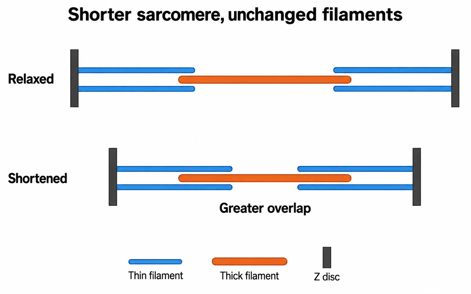
As the sarcomere shortens, its Z discs approach, thin-thick overlap increases, and the I band and H zone narrow. The A-band width remains essentially unchanged in this introductory sliding-filament model. The illustration is schematic: parallel coloured rods make overlap visible, but do not represent the full three-dimensional arrangement or the many myosin heads.
Contraction and shortening are not perfect synonyms
An activated muscle can develop tension without appreciable overall shortening, as when holding a load still. The diagrams explain a shortening sarcomere. They should not be read as a claim that every active muscle must visibly shorten or move a joint.
Measure the right structure
Predictions apply to shortening in the introductory sliding-filament model.
| Feature | During shortening | Why |
|---|---|---|
| Z-disc separation | Decreases | Defines sarcomere length |
| A-band width | Remains essentially unchanged | Tracks thick-filament length |
| I-band width | Decreases | Less thin-only region |
| H-zone width | Decreases and may disappear | Less central thick-only region |
| Individual filament lengths | Remain unchanged | Sliding changes overlap instead |
Use a ruler to reject the wrong mechanism
In an invented measurement, a sarcomere changes from 2.5 to 2.0 micrometres while the A band remains 1.6 micrometres wide. Sarcomere shortening is 0.5/2.5 x 100 = 20%. The unchanged A band supports unchanged thick-filament length. Reporting 'myosin shortened by 20%' would apply the percentage to the wrong structure.
The sarcomere lengths alone do not give the exact width of the H zone: additional information about thin-filament length and overlap is needed. Nor can one measurement determine whole-muscle force. This example is a geometry check, not a physiological dataset or a claim that every active muscle shortens by that fraction.
A useful drawing test is to keep every coloured filament the same length in both panels, move the boundaries closer, and increase overlap. If your drawing achieves shortening by erasing the ends of the filaments, it depicts the wrong mechanism even if the labels are correct.
The nerve signal opens access; calcium does not pull actin
At the neuromuscular junction, a motor-neuron signal leads to acetylcholine release and an electrical response in the muscle membrane. Excitation within the fibre triggers calcium release from the sarcoplasmic reticulum. The electrical event and the mechanical response are connected, but they are not identical events occurring at a single structure.
Calcium binds to troponin in the thin-filament regulatory system, changing the position of tropomyosin so that myosin-binding sites on actin become accessible. Calcium therefore permits cross-bridge activity. It is not a hook that directly drags a thin filament towards the centre, and it does not replace ATP as an energy source.
A motor unit comprises a motor neuron and the muscle fibres it supplies. It is a functional grouping, unlike a sarcomere, which is a repeating structural unit within a myofibril. Confusing those two units joins levels of organisation that should remain separate.
ATP is needed for release as well as repeated pulling
An energised myosin head can bind exposed actin. Changes in the head during the cross-bridge cycle pull the thin filament relative to the thick filament. A new ATP molecule binding to myosin allows detachment from actin. ATP hydrolysis then helps reset the head for another cycle. Saying only 'ATP makes contraction happen' hides the especially important detachment step.
For a reasoning check, suppose binding sites are accessible but no new ATP can bind to an attached head. The immediate textbook prediction is failure of normal detachment, not an extra-fast power stroke. This isolates one step conceptually; it is not an experiment to perform on a person or an explanation of every cause of muscle stiffness.
Relaxation also requires calcium to be removed from the sarcoplasm, mainly by pumping it back into the sarcoplasmic reticulum. As regulatory sites lose calcium, access to actin is restricted again. ATP supports calcium pumping as well as cross-bridge cycling, so relaxation should not be described as a process requiring no energy anywhere.
Striated does not automatically mean voluntary
Skeletal muscle is striated and generally under voluntary control, although reflex activity also uses it. Cardiac muscle is striated but involuntary. Smooth muscle lacks the same visible banding pattern and is involuntary, with roles in the walls of hollow organs. Appearance and control are separate classification axes: one cannot be deduced reliably from the other.
Other movement mechanisms also belong in this chapter. Amoeboid movement uses changing cell shape and pseudopodia; cilia can move material along an epithelial surface. These movements should not be explained by sarcomeres simply because both involve motion. The appropriate structural mechanism depends on the cell or tissue involved.
For retrieval, place heart muscle and skeletal muscle beside each other and state one shared feature and one difference. Both are striated; their control and cellular organisation differ. That comparison is more useful than treating 'striated' and 'skeletal' as interchangeable words.
Bones and joints turn fibre activity into a body movement
Skeletal muscles exert pulls through their attachments; opposite movements at a joint commonly use opposing muscle groups. A flexor and an extensor are named for their actions, not because one is always active and the other always inactive. The joint and arrangement of attachments constrain the movement that a pull can produce.
Fibrous, cartilaginous and synovial joints differ in their connections and permitted movement. A hinge permits movement mainly in one plane, whereas a ball-and-socket joint allows movement in several planes. Those descriptions concern joint geometry, not a different biochemical ATP cycle for each joint. The axial skeleton and appendicular skeleton likewise classify location, not muscle-control type.
To reconstruct the complete explanation, trace motor-neuron signal, muscle excitation, calcium availability, cross-bridge cycling, sarcomere shortening and joint movement. Then point out where ATP participates and which length stays constant. NCERT anchor: Locomotion and Movement, sections 17.2.1-17.2.2 and 17.4 in the 2025-26 reprint. This is an educational mechanism guide, not advice for investigating pain or treating a movement disorder.
Common confusions to check
- An A band includes overlap; it is not all thick-only territory.
- Cardiac muscle is striated but involuntary.
- ATP binding permits detachment rather than permanently fixing the cross-bridge.
Editorial note and disclaimer
Written by: DongFeng. Published by: MedQGo. Last updated: September 23, 2026.
Disclaimer: This guide is a revision aid for NEET-UG aspirants and does not constitute medical advice. For clinical or health-related queries, consult a qualified medical professional.
References
Related revision guides
How to use this guide
Read the relevant NCERT chapter first. Then redraw the relationships or process described here from memory, compare your version with the textbook, and correct only the gaps. This is an independent revision aid, not official NCERT, NTA, or NEET material.