Molecular analysis asks a sequence of different questions
A DNA sample is not automatically ready to answer every laboratory question. A revision-friendly route is: obtain a DNA-containing sample, focus on a chosen sequence when needed, make enough copies for observation, and separate fragments when their sizes need to be compared. The method changes because the question changes.
Restriction enzymes, PCR and gel electrophoresis often appear together, but they do not do the same job. A restriction endonuclease recognises particular DNA sequences and cuts DNA. PCR amplifies a selected region. Gel electrophoresis separates DNA fragments after an electric field moves them through a gel. Say the verb beside each tool before choosing an option.
PCR copies a target; it does not sort fragments
Polymerase chain reaction is designed to amplify a selected DNA region. In each cycle, double-stranded DNA is separated, primers bind to complementary target sequences, and a DNA polymerase extends from the primers. Repeating the cycle produces many copies of the region defined by the primers.
The distinction matters because PCR is about increasing the amount of a chosen sequence, whereas a gel is about separating molecules already present in a sample. A primer is not the enzyme that copies DNA, and a thermal cycle is not a step of gel electrophoresis. Track the input and output: target DNA enters PCR; more copies of that target leave it.
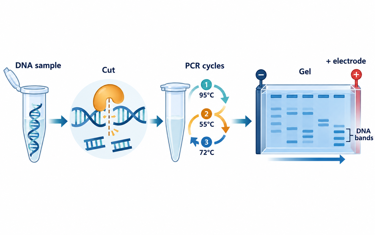
The cut-copy-separate check
Before naming a technique, identify whether the sample needs to be changed, amplified or compared by size.
- Restriction endonucleases cut DNA at recognised sequences.
- PCR uses repeated denaturation, primer binding and extension to amplify a defined region.
- DNA migrates toward the positive electrode in an agarose gel because its phosphate backbone is negatively charged.
- Smaller DNA fragments generally move farther through the gel during the same run.
Do not let three laboratory verbs collapse into one
Cutting, copying and separating can happen in the same broader investigation, but each answers a different question. A restriction enzyme changes a DNA molecule at a recognition sequence; PCR raises the number of copies of a selected region; an electric field separates fragments in a gel.
When an option combines a correct tool with the wrong outcome, return to the sample. Did it become fragments, many copies, or visible bands? That single check usually restores the method.
DNA analysis: match the question to the tool
A method is easier to identify when you name the change it makes to the sample or the observation it produces.
| Question about the sample | Useful tool or step | What the output shows | Nearby but different tool |
|---|---|---|---|
| Where can DNA be cut? | Restriction endonuclease | Fragments formed at recognised sequences | DNA ligase, which joins fragments |
| How can one target region be made plentiful? | PCR | Many copies of a primer-defined DNA region | Gel electrophoresis, which separates fragments |
| How can fragment sizes be compared? | Gel electrophoresis | Band positions after migration through a gel | PCR, which amplifies rather than sorts |
| Which way does DNA move in an agarose gel? | Electric field | Toward the positive electrode | Movement toward the negative electrode |
A gel turns fragment size into a visible pattern
DNA carries a negative charge because of its phosphate backbone. In an agarose gel, DNA fragments move toward the positive electrode when an electric field is applied. The gel acts as a molecular sieve, so smaller fragments generally travel farther through it than larger fragments during the same run.
A lane is one sample path; a band marks DNA fragments that have travelled a similar distance. The migration pattern can help compare fragment sizes, but it does not provide the DNA sequence merely by looking at a band. Before interpreting a gel, check the direction of travel, the sample wells, and whether the prompt asks about size, copying, cutting, or detection.
Build a tool-to-output map from memory
Draw three boxes labelled cut, copy and separate. Put a restriction endonuclease above the first box, PCR above the second, and agarose gel electrophoresis above the third. Under each, write one output: fragments, many target copies, and separated bands. This turns a vocabulary list into a usable decision map.
Then add a fourth box labelled detect a specific sequence. This reminds you that a gel pattern alone and a sequence-specific identification are different levels of analysis. The point is not to memorise every laboratory technique beyond the chapter; it is to protect the role of each NCERT-linked tool.
Common confusions to check
- PCR amplifies a target sequence; it does not separate DNA fragments by size.
- DNA ligase joins fragments, whereas a restriction endonuclease cuts DNA at recognised sequences.
- DNA is negatively charged and migrates toward the positive electrode in a gel.
Editorial note and disclaimer
Written by: DongFeng. Published by: MedQGo. Last updated: September 20, 2026.
Disclaimer: This guide is a revision aid for NEET-UG aspirants and does not constitute medical advice. For clinical or health-related queries, consult a qualified medical professional.
References
Related revision guides
How to use this guide
Read the relevant NCERT chapter first. Then redraw the relationships or process described here from memory, compare your version with the textbook, and correct only the gaps. This is an independent revision aid, not official NCERT, NTA, or NEET material.