Build every plant section from three tissue systems
Plant anatomy asks where tissues are placed and how that arrangement supports function. Begin with three systems. Dermal tissue forms the outer boundary, ground tissue fills or differentiates much of the interior, and vascular tissue conducts water, minerals and organic solutes. A root, stem and leaf contain these systems in different spatial arrangements.
Read a transverse section from outside inward. First locate the epidermal boundary, then identify cortex or other ground tissue, and finally find the vascular region. This outside-to-inside route is more reliable than searching immediately for a single memorised shape, because the same tissue name can occupy a different geometry in another organ.
- Dermal system: protection and exchange at the outer surface.
- Ground system: photosynthesis, storage and mechanical support depending on organ and cell type.
- Vascular system: xylem and phloem arranged as bundles or a central vascular cylinder.
Meristematic and permanent tissues answer different questions
Meristematic cells retain the capacity for division. Apical meristems extend roots and shoots, intercalary meristems contribute to growth at bases or nodes in grasses, and lateral meristems increase girth. Permanent tissues are differentiated for particular roles and are classified as simple or complex according to their cellular composition.
Parenchyma cells are generally living and thin-walled, with roles in storage, photosynthesis and repair. Collenchyma provides flexible support through uneven wall thickening and is commonly associated with growing parts. Sclerenchyma has thick, lignified walls and provides mechanical strength; its cells are usually dead at maturity. The useful comparison is wall character plus living state plus function, not hardness alone.
Do not identify a section from one isolated feature
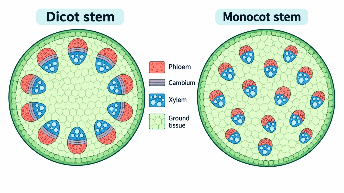
A large pith, an open bundle or an exarch xylem pattern is useful only when placed beside the rest of the section. Root-versus-stem identity comes first from vascular geometry; dicot-versus-monocot identity is then strengthened by xylem number, pith, bundle arrangement and cambium.
The same discipline applies to tissues. Xylem is not entirely dead because its parenchyma is living, and phloem is not entirely living because its fibres are dead. A precise answer names the component rather than extending one property to the whole complex tissue.
Four transverse sections: the clues that identify them
Start with vascular arrangement, then check cambium and pith. These axes separate root from stem and dicot from monocot without relying on one memorised picture.
| Section | Vascular arrangement | Pith and cambium | Decisive distinction |
|---|---|---|---|
| Dicot root | Radial; few xylem arms alternate with phloem | Pith small or inconspicuous; no bundle cambium pattern | Central star-like xylem with exarch development |
| Monocot root | Radial and polyarch | Large central pith | Many xylem groups around the pith |
| Dicot stem | Conjoint collateral bundles in a ring | Open bundles with cambium; distinct pith | Ring arrangement supports secondary growth |
| Monocot stem | Conjoint collateral bundles scattered in ground tissue | Closed bundles; no distinct central pith | Bundle sheath and scattered distribution |
Xylem and phloem are complex tissues with unequal living states
Xylem conducts water and minerals and also contributes support. Tracheids and vessel elements are the principal conducting elements and are dead at maturity; xylem fibres are also dead, while xylem parenchyma is living. Phloem transports organic material. Sieve-tube elements work with companion cells in angiosperms, while phloem parenchyma and fibres complete the tissue described in the chapter.
Avoid the shortcut that every xylem element is dead or every phloem element is living. Xylem parenchyma is the living exception within xylem, and phloem fibres are dead. In a vascular bundle, xylem usually lies toward the inner side and phloem toward the outer side in a stem, but roots arrange the tissues on different radii rather than as the same collateral bundle.
Root sections: use xylem geometry and pith as anchors
A young root has an epiblema or piliferous layer, cortex, endodermis, pericycle and a central vascular region. The endodermis forms the innermost cortical layer, while the pericycle lies just inside it and can give rise to lateral roots. Xylem and phloem occur on alternate radii, a radial arrangement that distinguishes the root from a typical stem bundle.
In a typical dicot root, central xylem forms relatively few arms and the pith is small or inconspicuous. In a monocot root, xylem is polyarch and a large pith occupies the centre. Both can show an exarch xylem condition, with protoxylem toward the outside. Therefore the presence of radial bundles alone identifies a root; the number of xylem arms and the size of the pith help separate the two root types.
Stem sections: ring versus scattered bundles is the first split
A dicot stem typically shows a distinct cortex, vascular bundles arranged in a ring and a central pith. The bundles are conjoint and collateral, with phloem outside and xylem inside. Cambium lies between them in an open bundle, allowing the possibility of secondary growth.
A monocot stem has numerous vascular bundles scattered through undifferentiated ground tissue. The bundles are conjoint, collateral and closed because vascular cambium is absent within them. Each bundle has a sclerenchymatous sheath, and a protoxylem lacuna may be visible. Scattered does not mean disorganised; bundle size and density can vary from the outer to inner ground tissue.
The clean diagnostic order is therefore arrangement, cambium and ground-tissue differentiation. Starting with a single vessel diameter can mislead because section quality and age alter visual detail.
Leaf anatomy links surface, mesophyll and vascular orientation
A dorsiventral dicot leaf has upper and lower epidermis with mesophyll differentiated into palisade and spongy regions. Stomata are usually more numerous on the lower surface. Vascular bundles run through the mesophyll, and xylem faces the upper, adaxial side while phloem faces the lower, abaxial side.
An isobilateral monocot leaf has less sharply differentiated mesophyll and commonly shows stomata on both surfaces. Bulliform cells in the upper epidermis are associated with leaf rolling under water stress in grasses. Parallel venation at the morphological level corresponds with multiple vascular bundles seen in section, but venation and internal tissue arrangement remain different observations.
Secondary growth changes girth by adding tissues in rings
In a typical dicot stem, interfascicular and intrafascicular cambial regions form a continuous vascular cambium ring. Cambium produces secondary xylem toward the inside and secondary phloem toward the outside. Because much more secondary xylem accumulates, the woody region expands inward from the cambial boundary.
Cork cambium develops in outer tissues and forms protective derivatives as the original epidermis can no longer accommodate increasing girth. Annual-ring interpretation belongs to seasonal differences in secondary xylem, not to a new set of primary vascular bundles. Keep this overview at the chapter level; detailed wood identification is outside the revision goal.
An outside-in transverse-section recall routine
Make four empty circles labelled dicot root, monocot root, dicot stem and monocot stem. In each, place xylem and phloem before adding cortex or pith. Then mark radial versus conjoint arrangement, ring versus scattered bundles, and open versus closed bundles. The finished page should let another learner identify each section from three visible clues.
Finish by drawing one leaf rectangle with upper and lower surfaces. Add palisade tissue, spongy tissue and a vascular bundle with xylem above phloem. Say aloud why this is a dorsiventral leaf, then list the two features you would change for an isobilateral monocot leaf.
Common confusions to check
- Xylem parenchyma is living even though the main xylem conducting elements are dead at maturity.
- Roots have radial xylem and phloem, whereas typical stem bundles are conjoint and collateral.
- Dicot stem bundles are commonly arranged in a ring and open; monocot stem bundles are scattered and closed.
Editorial note and disclaimer
Written by: DongFeng. Published by: MedQGo. Last updated: September 21, 2026.
Disclaimer: This guide is a revision aid for NEET-UG aspirants and does not constitute medical advice. For clinical or health-related queries, consult a qualified medical professional.
References
Related revision guides
How to use this guide
Read the relevant NCERT chapter first. Then redraw the relationships or process described here from memory, compare your version with the textbook, and correct only the gaps. This is an independent revision aid, not official NCERT, NTA, or NEET material.